Short lessons, one track at a time
Five courses of four short video lessons, each built around one idea you can use at the bedside. Pick the track that matches your work and take the lessons in order. Until the videos are ready, every lesson links to a simulator, case or page where you can practise it today.
Recording now with Dr. Nassef- 5tracks, one for each kind of clinical work
- 20short video lessons, four per track, in order
- Freeopen teaching; not CME-accredited
- ForMedical students, residents, ICU, emergency, anaesthesia, paediatric and neonatal teams
 Track 1 of 5
4 video lessons
Foundations
For medical studentsWhy blood pressure is not flow, and the three levers that set stroke volume.
What you’ll be able to do
- Explain why a normal blood pressure does not prove normal flow
- Predict how preload, afterload and contractility change stroke volume
- Read indexed values instead of raw numbers
-
 Coming soon
Lesson 1 of 4: video coming soon
Flow is not pressure
Why blood pressure can look normal while flow is already falling: pressure is flow times resistance.
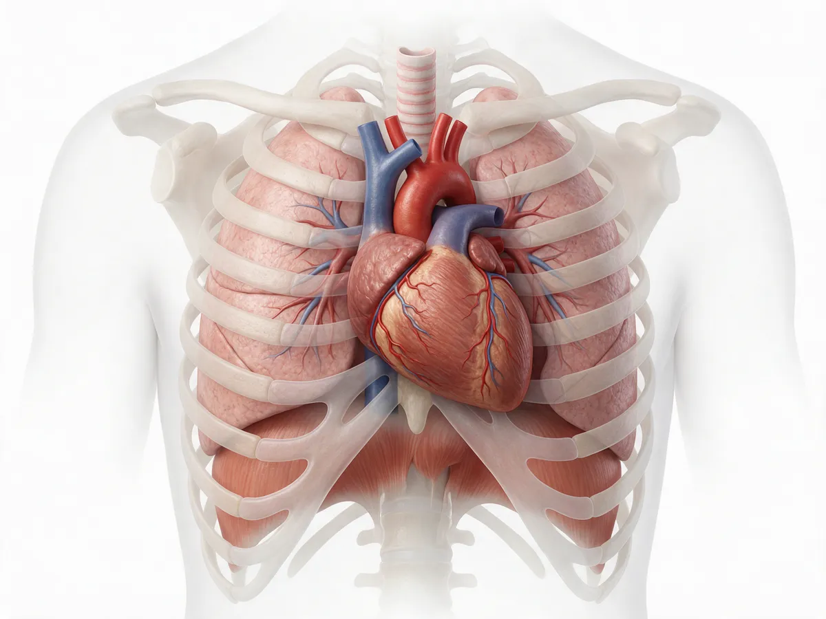
Anatomy basics Practise nowBleed a patient and see which monitor notices first -
 Coming soon
Lesson 2 of 4: video coming soon
Preload, afterload, contractility
The three levers that set stroke volume: how full the heart is, what it pushes against, and how hard it squeezes.
Anatomy basics Practise nowMove the three levers in the simulator -
 Coming soon
Lesson 3 of 4: video coming soon
Frank–Starling in one picture
On the steep part of the curve, filling raises stroke volume; on the flat part it adds little except congestion.
Practise nowFind the flat part of the curve -
 Coming soon
Lesson 4 of 4: video coming soon
Indexing to body size
Why stroke index and cardiac index let you compare patients of any size, from a newborn to a large adult.
Practise nowRead the indexed parameters
 Track 2 of 5
4 video lessons
Shock states
For residentsTell the four kinds of shock apart from their hemodynamic fingerprint.
What you’ll be able to do
- Name the likely mechanism from a hemodynamic profile
- Match the first treatment to that mechanism
- Spot mixed pictures that need bedside imaging to confirm
-
 Coming soon
Lesson 1 of 4: video coming soon
Hypovolemic
Low stroke volume, high resistance and a dry chest: an empty tank, with a stroke volume that rises on a leg raise.
Practise nowCase: vomiting, and still on a diuretic -
 Coming soon
Lesson 2 of 4: video coming soon
Distributive and septic
Very low resistance, often with a high output once filled: vessel tone, not only filling, drives the low pressure.
Practise nowCase: fever, confusion and a low pressure -
Coming soon
Lesson 3 of 4: video coming soon
Cardiogenic
Low output and low contractility with normal or high resistance: a weak pump, not an empty tank.
Practise nowCase: cold, grey and hypotensive -
 Coming soon
Lesson 4 of 4: video coming soon
Reading the pattern
Put stroke volume, resistance, contractility and thoracic fluid together to name the mechanism, obstructive shock included.
Practise nowCompare the shock fingerprints
 Track 3 of 5
4 video lessons
Fluid responsiveness
For ICU and emergencyGive fluid to those who will respond, and stop before it harms.
What you’ll be able to do
- Decide who needs a fluid-responsiveness test
- Run and read a passive leg raise
- Know when stroke volume variation can and cannot be used
- Recognise when to stop giving fluid
-
 Coming soon
Lesson 1 of 4: video coming soon
Who will respond?
Only about half of unstable patients raise their output with fluid, so test before you give.
Practise nowTest the response to filling in the simulator -
Coming soon
Lesson 2 of 4: video coming soon
Passive leg raise
A reversible fluid challenge from the patient’s own legs: a stroke volume rise of about 10% or more predicts a response.
Practise nowCase: do the leg raise yourself -
Coming soon
Lesson 3 of 4: video coming soon
Stroke volume variation
What a high SVV means, and why it misleads with spontaneous breathing, small tidal volumes or an irregular rhythm.
Practise nowCase: when SVV cannot be trusted -
Coming soon
Lesson 4 of 4: video coming soon
When to stop
Stop when stroke volume no longer rises and thoracic fluid starts to climb: the flat part of the curve.
The fluid decision rule Practise nowCase: when more fluid would harm
 Track 4 of 5
4 video lessons
Paediatric and neonatal
For paediatric and neonatal teamsChildren are not small adults: their own numbers, their own shock.
What you’ll be able to do
- Read a child’s numbers against indexed, age-specific values
- Recognise low-output septic shock in a child
- Follow the trend, not one pressure, in a preterm baby
-
Coming soon
Lesson 1 of 4: video coming soon
Not small adults
More flow per kilogram, faster hearts and different compensation: why adult numbers mislead in children.
Practise nowCompare newborn and adult flow -
Coming soon
Lesson 2 of 4: video coming soon
Indexed normal values
Never compare raw values across ages: read each child against indexed, age-specific ranges and their own trend.
Practise nowSee which values are indexed -
Coming soon
Lesson 3 of 4: video coming soon
Septic shock in children
Children with septic shock more often have low output and high resistance than adults do: measure, don’t assume.
Practise nowSee how septic shock can turn low-output -
Coming soon
Lesson 4 of 4: video coming soon
The preterm circulation
Why a steady blood pressure in a preterm baby can hide falling flow, and what the trend adds to echocardiography.
Practise nowCase: a 28-week preterm on day two
 Track 5 of 5
4 video lessons
Non-invasive monitoring
For anaesthesia and ICUFrom four skin sensors to a number you can trust, and when not to.
What you’ll be able to do
- Explain Electrical Cardiometry in plain words
- Place the four sensors correctly
- Read every number on the monitor screen
- Know when to trust the trend, and when not to
-
Coming soon
Lesson 1 of 4: video coming soon
How Electrical Cardiometry works
From a tiny current across the chest to a stroke volume, beat by beat, without a catheter.
Practise nowSee one heartbeat, slowed down -
Coming soon
Lesson 2 of 4: video coming soon
Placing the four sensors
Where the four sensors go on the left side, and why moving them off position distorts the signal.
Practise nowPlace the four sensors -
Coming soon
Lesson 3 of 4: video coming soon
Reading the screen
What each number means: flow, resistance, contractility and thoracic fluid, and their indexed forms.
Practise nowEvery parameter, in plain words -
Coming soon
Lesson 4 of 4: video coming soon
Trusting the trend
Why single values carry a wide error while changes after a leg raise, fluid or a drug are more reliable, and when to trust neither.
The evidence Practise nowWhen to be careful with the numbers
Watch one idea. Use it. Check yourself.
Every lesson follows the same three steps. The second and third already work today.
-
1. Watch a short video
Dr. Nassef explains one idea per lesson, with the monitor and the physiology side by side.
Being recorded -
2. Practise it
Move the levers in the simulator, or work through the matching page, and watch the numbers answer.
Available today -
3. Check yourself
Each interactive case asks for your decision first, then shows the reasoning and the teaching point.
Available today
The courses are free. They are teaching, not accreditation: the academy is not CME-accredited and does not issue certificates. Clinical content is under review by Dr. Nassef.
Start with the simulator today
Load a septic, hypovolemic or cardiogenic patient, move heart rate, filling, contractility and resistance, and see stroke volume, cardiac output and pressure answer on the Frank–Starling curve.